DESCRIPTION
The tissue culture shown in this scanning electron microscope image is infected with the Human Immunodeficiency Virus, or HIV. • SIZE: HIV particles are 90-120 nm in diameter. • IMAGING TOOL: Scanning electron microscope
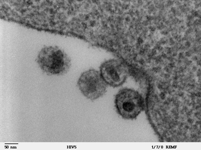
DESCRIPTION
The tissue culture shown in this scanning electron microscope image is infected with the Human Immunodeficiency Virus, or HIV. • SIZE: HIV particles are 90-120 nm in diameter. • IMAGING TOOL: Scanning electron microscope
DOWNLOAD FILES
Credits
Dartmouth Electron Microscope Facility - Attribution is required. The creator listed here has made this image available to NISE Network partners for non-profit educational use only. Uses may include but are not limited to reproduction and distribution of copies, creation of derivative works, and combination with other assets to create exhibitions, programs, publications, research, and websites.
The creator listed above has made this image available to NISE Network partners for non-profit educational use only. Uses may include but are not limited to reproduction and distribution of copies, creation of derivative works, and combination with other assets to create exhibitions, programs, publications, research, and websites.