DESCRIPTION
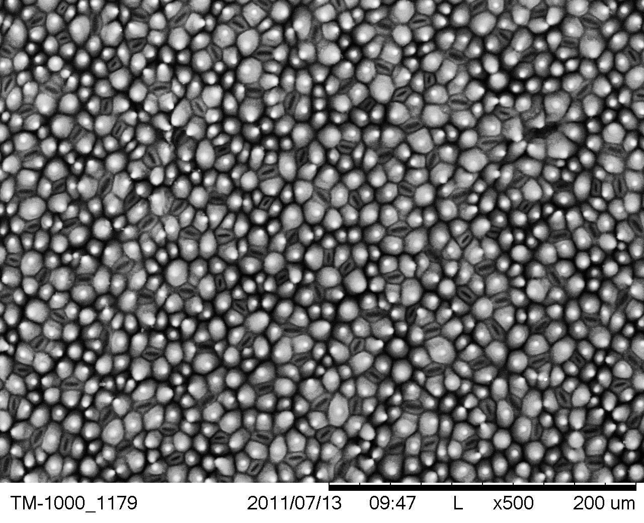
Scanning electron microscope image of the surface of a lotus leaf. • SIZE: Scale bar representes 200 µm • IMAGING TOOL: Table-top Scanning Electron Microscope (SEM)
DESCRIPTION
Scanning electron microscope image of the surface of a lotus leaf. • SIZE: Scale bar representes 200 µm • IMAGING TOOL: Table-top Scanning Electron Microscope (SEM)
Credits
Rashmi Nanjundaswamy / Lawrence Hall of Science
Developed for the NISE Network with funding from the National Science Foundation under Award Numbers 0532536 and 0940143. Any opinions, findings, and conclusions or recommendations expressed in this product are those of the authors and do not necessarily reflect the views of the Foundation.
Creative Commons Attribution Non-Commercial Share Alike 3.0 United States (CC BY-NC-SA 3.0 US).
View more details

NISE Network products are developed through an iterative collaborative process that includes scientific review, peer review, and visitor evaluation in accordance with an inclusive audiences approach. Products are designed to be easily edited and adapted for different audiences under a Creative Commons Attribution Non-Commercial Share Alike license. To learn more, visit our Development Process page.

